Welcome to the Amira-Avizo Software Use Case Gallery
Below you will find a collection of use cases of our 3D data visualization and analysis software. These use cases include scientific publications, articles, papers, posters, presentations or even videos that show how Amira-Avizo Software is used to address various scientific and industrial research topics.
Use the Domain selector to filter by main application area, and use the Search box to enter keywords related to specific topics you are interested in.
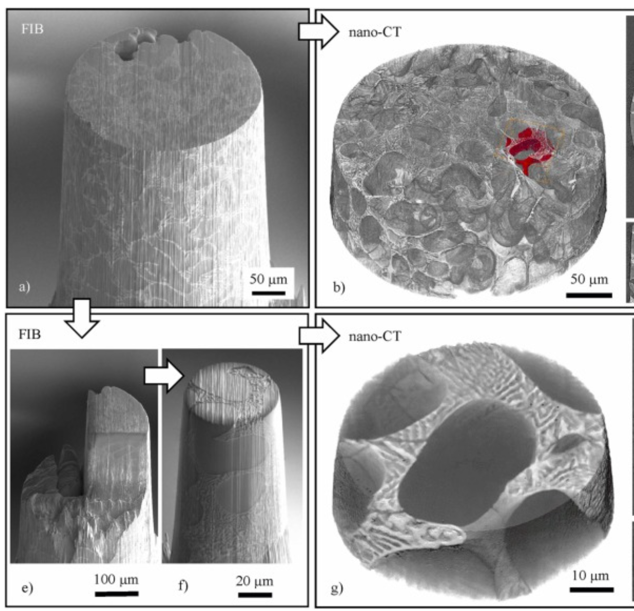
Three-dimensional imaging of microstructural evolution in SEM-based nano-CT
Scanning electron microscopy (SEM) is a powerful and versatile technique for materials characterization and present in many laboratories. The integration of an X-ray target holder and detector allows expanding the modalities of SEM by X-ray imaging. These little hardware adaptations enable radiography ... Read more
Jonas Fell, Christoph Pauly, Michael Maisl, Simon Zabler, Frank Mücklich, Hans-Georg Herrmann
Recycling spent lithium-ion batteries (LiBs) guarantees the conservation of important metal resources by reducing demands on raw supply and offsetting the energy and environmental costs associated with its manufacture. Employing a molten salt as a solvent for extraction affords a much greener and simpler route to metal recovery by electrochemical means. The current mechanistic understanding of the electrochemical recovery of metals in molten salts needs to be improved for the process to be op... Read more
Mateen Mirza, Wenjia Du, Lara Rasha, Steven Wilcock, Arfon H. Jones, Paul R. Shearing, Dan J.L. Brett
The complex mechanical response of open-cell foams depends strongly on the hierarchy of length scales inherent in them, from engineering-part scale to the ligament scale through the grain scale down to the crystal-lattice scale. A first step toward understanding and predicting the coordinated mechanical response across le... Read more
Jayden C. Plumb, Jonathan F. Lind, Joseph C. Tucker, Ron Kelley, Ashley D. Spear
The usefulness of desktop Micro-CT scanners for the study of archaeological artefacts is demonstrated in a non-destructive study of manufacturing methods of Roman and Early Medieval monochrome and polychrome glass beads. Differences in glass colours show up in these scans as differences in attenuation. The presence and distribution of bubbles and various inclusions (metal, opacifier) are also well visible. Shaft shapes and patterns of bubbles inside the glass make it possible in most cases to... Read more
D.J.M.Ngan-Tillard, D.J.Huisman, F.Corbella, A.Van Nass
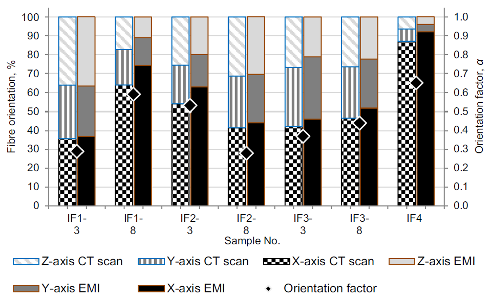
The interest in potential applications produced with self-compacting fibre reinforced concrete continues to grow, but in practice, problems associated with an uneven distribution and orientation of fibres in the concrete structure occur. It is not clear what exactly influences uneven distribution of fibres in selfcompacting concrete (SCC) mixtures, especially during the casting and how different factors influence fibre orientation. The objective of this work was to investigate how rheological... Read more
Elena Jasiuniene, Vaidotas Cicenas, Paulius Grigaliunas, Zymantas Rudzionis, Arunas Aleksandras Navickas
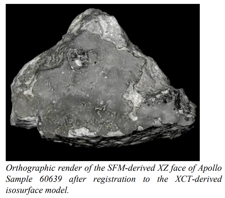
Our team is developing a modern, cross-disciplinary approach to documentation and preservation of astromaterials, specifically lunar and meteorite samples stored at the Johnson Space Center (JSC) Lunar Sample Laboratory Facility.
Apollo Lunar Sample 60639, collected as part of rake sample 60610 during the 3rd Extra-Vehicular Activity of the Apollo 16 mission in 1972, served as the first NASA preserved lunar sample to be examined by our team in the development of a novel approach to int... Read more
K.R. Beaulieu , E.H. Blumenfeld , D.A. Liddle , E.R. Oshel , C.A. Evans , R.A. Zeigler , K. Righter , R.D. Hanna , R.A. Ketcham
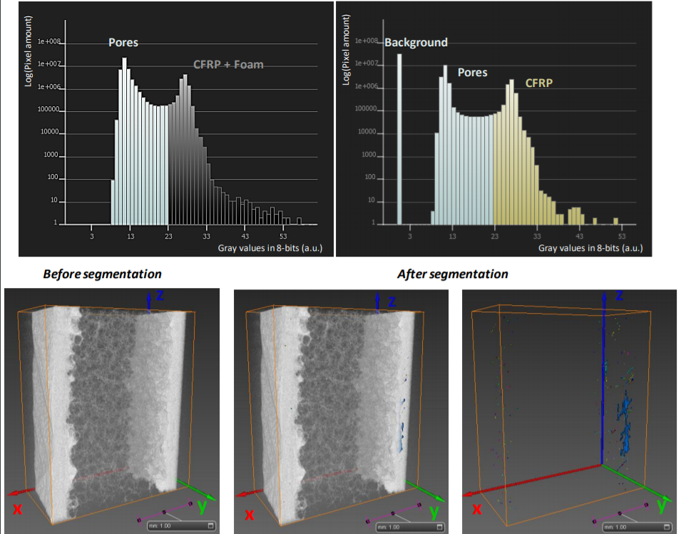
The aim of the current study is to propose a versatile, non-destructive inspection strategy to evaluate the structure of two different aircraft carbon fibre reinforced polymer (CFRP) -based composite configurations, which are widely used for structural elements, respectively layered composite and sandwich structure. X-ray computed tomography (CT) has been used as a flexible method for assessment of porosity levels in CFRP components in both types of configuration, permitting to investigate th... Read more
Elena Dilonardo, Michele Nacucchi, Fabio De Pascalis, Mauro Zarrelli, Cinzia Giannini
Microstructural analysis of TRISO particles using multi-scale X-ray computed tomography
TRISO particles, a composite nuclear fuel built up by ceramic and graphitic layers, have outstanding high temperature resistance. TRISO fuel is the key technology for High Temperature Reactors (HTRs) and the Generation IV Very High Temperature Reactor (VHTR) variant.
TRISO offers unparalleled containment of fission products and is extremely robust during accident conditions. An understanding of the thermal performance and mechanical properties of TRISO fuel requires a detailed knowledg... Read more
T. Lowe, R.S. Bradley, S. Yue, K. Barii, J. Gelb, N. Rohbeck, J. Turner, P.J. Withers
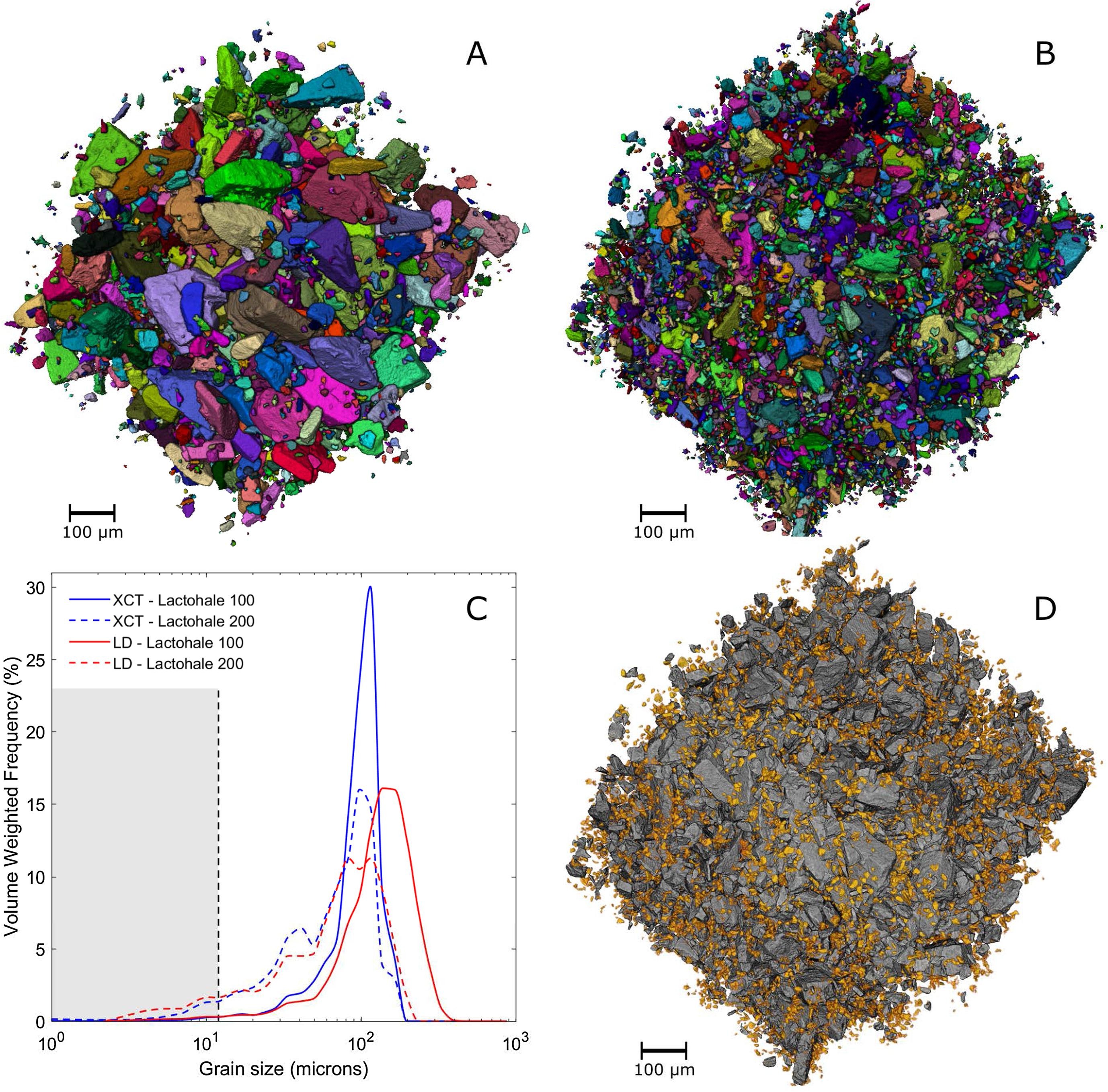
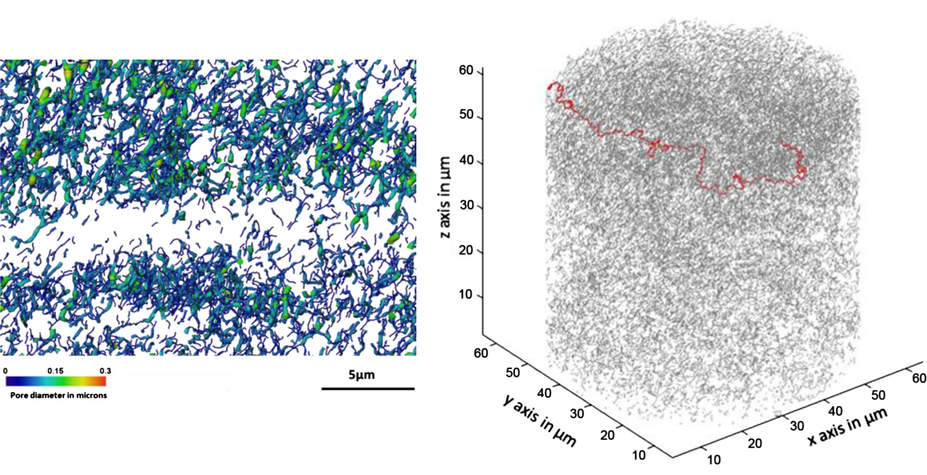
3D characterisation of dry powder inhaler formulations
Carrier-based dry powder inhaler (DPI) formulations need to be accurately characterised for their particle size distributions, surface roughnesses, fines contents and flow properties. Understanding the micro-structure of the powder formulation is crucial, yet current characterisation methods give incomplete information. Commonly used techniques like laser diffraction (LD) and optical microscopy (OM) are limited due to the assumption of sphericity and can give variable results depending on par... Read more
P. Gajjar, I.D. Styliari, T.T.H. Nguyen, J. Carr, X. Chen, J.A. Elliott, R.B. Hammond, T.L. Burnett, K. Roberts, P.J. Withers, D.Murnane
Computed tomography is an increasingly popular technique for the non-destructive study of fossils. Whilst the science of X-ray computed tomography (CT) has greatly matured since its first fossil applications in the early 1980s, the applications and limitations of neutron tomography (NT) remain relatively unexplored in palaeontology. These highest resolution neutron tomographic scans in palaeontology to date were conducted on a specimen of Austrosequoia novae-zeelandiae (Ettingshausen) Mays an... Read more
Chris Mays, Joseph J. Bevitt, and Jeffrey D. Stilwell
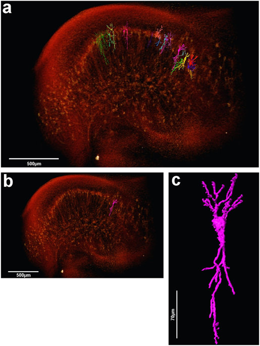
The assessment of neuronal number, spatial organization and connectivity is fundamental for a complete understanding of brain function. However, the evaluation of the three-dimensional (3D) brain cytoarchitecture at cellular resolution persists as a great challenge in the field of neuroscience. In this context, X-ray microtomography has shown to be a valuable non-destructive tool for imaging a broad range of samples, from dense materials to soft biological specimens, arisen as a new method fo... Read more
Matheus de Castro Fonseca, Bruno Henrique Silva Araujo, Carlos Sato Baraldi Dias, Nathaly Lopes Archilha, Dionísio Pedro Amorim Neto, Esper Cavalheiro, Harry Westfahl Jr, Antônio José Roque da Silva, Kleber Gomes Franchini
Ptychographic X-ray computed tomography (PXCT) is a quantitative imaging modality that non-destructively maps the 3D electron density inside an object with tens of nanometers spatial resolution. This method provides unique access to the morphology and structure of the osteocyte lacuno-canalicular network (LCN) and nanoscale density of the tissue in the vicinity of an osteocyte lacuna. Our findings indicate that PXCT can non-destructively provide detailed, nanoscale information on the 3D org... Read more
Antonia Ciani, Hechmi Toumi, Stéphane Pallu, Esther H.R.Tsai, Ana Diaz, Manuel Guizar-Sicairos, Mirko Holler, Eric Lespessailles, Cameron M.Kewish | Synchrotron Soleil, France

Investigation of Carbon Fiber Architecture in Braided Composites Using X-Ray CT Inspection
During the fabrication of braided carbon fiber composite materials, process variations occur which affect the fiber architecture.
Quantitative measurements of local and global fiber architecture variations are needed to determine the potential effect of process variations on mechanical properties of the cured composite. Although non-destructive inspection via X-ray CT imaging is a promising approach, difficulties in quantitative analysis of the data arise due to the similar densities o... Read more
Daniel J. Rhoads, Sandi G. Miller, Gary D. Roberts, Richard W. Rauser, Dmitry Golovaty, J. Patrick Wilber, Malena I. Español
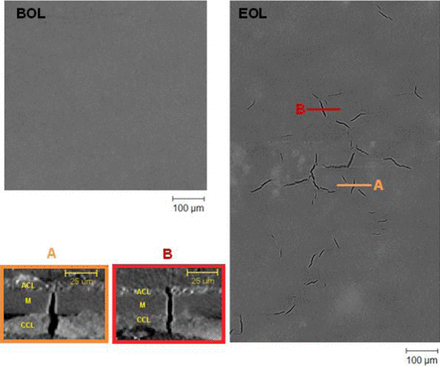
3D Failure Analysis of Pure Mechanical and Pure Chemical Degradation in Fuel Cell Membranes
Lifetime-limiting failure of fuel cell membranes is generally attributed to their chemical and/or mechanical degradation. Although both of these degradation modes occur concurrently during operational duty cycles, their uncoupled investigations can provide useful insights into their individual characteristics and consequential impacts on the overall membrane failure.
X-ray computed tomography is emerging as an advantageous tool for fuel cell failure analysis due to its non-destructive ... Read more
Yadvinder Singh, Francesco P. Orfino, Monica Dutta, and Erik Kjeang
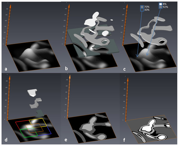
MIA-Clustering: a novel method for segmentation of paleontological material
Paleontological research increasingly uses high-resolution micro-computed tomography (μCT) to study the inner architecture of modern and fossil bone material to answer important questions regarding vertebrate evolution. This non-destructive method allows for the measurement of otherwise inaccessible morphology. Digital measurement is predicated on the accurate segmentation of modern or fossilized bone from other structures imaged in μCT scans, as errors in segmentation can result in inaccur... Read more
Christopher J. Dunmore, Gert Wollny, Matthew M. Skinner